
Page 553 - Atlas of Histology with Functional Correlations
P. 553
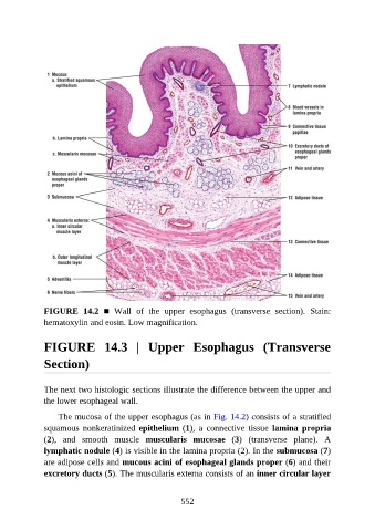
FIGURE  14.2  ■  Wall  of  the  upper  esophagus  (transverse  section).  Stain:
               hematoxylin and eosin. Low magnification.


               FIGURE  14.3  |  Upper  Esophagus  (Transverse

               Section)


               The next two histologic sections illustrate the difference between the upper and

               the lower esophageal wall.

                   The mucosa of the upper esophagus (as in Fig. 14.2) consists of a stratified
               squamous  nonkeratinized  epithelium (1),  a  connective  tissue  lamina  propria
               (2),  and  smooth  muscle  muscularis  mucosae  (3)  (transverse  plane).  A

               lymphatic nodule (4) is visible in the lamina propria (2). In the submucosa (7)
               are adipose cells and mucous acini of esophageal glands proper (6) and their
               excretory ducts (5). The muscularis externa consists of an inner circular layer


                                                          552