
Page 723 - Atlas of Histology with Functional Correlations
P. 723
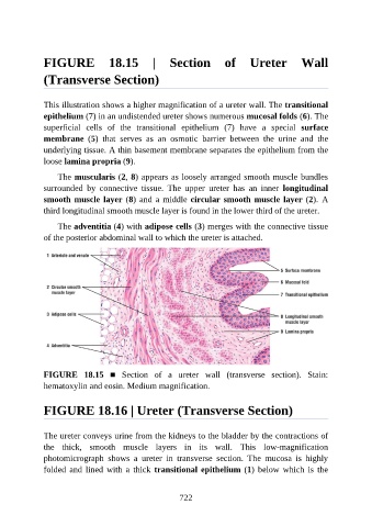
FIGURE  18.15  |  Section  of  Ureter  Wall

               (Transverse Section)


               This illustration shows a higher magnification of a ureter wall. The transitional
               epithelium (7) in an undistended ureter shows numerous mucosal folds (6). The

               superficial  cells  of  the  transitional  epithelium  (7)  have  a  special  surface
               membrane  (5)  that  serves  as  an  osmotic  barrier  between  the  urine  and  the
               underlying tissue. A thin basement membrane separates the epithelium from the

               loose lamina propria (9).

                   The muscularis (2, 8) appears as loosely arranged smooth muscle bundles
               surrounded  by  connective  tissue.  The  upper  ureter  has  an  inner  longitudinal
               smooth muscle layer (8) and a middle circular smooth muscle layer (2).  A
               third longitudinal smooth muscle layer is found in the lower third of the ureter.

                   The adventitia (4) with adipose cells (3) merges with the connective tissue

               of the posterior abdominal wall to which the ureter is attached.


               FIGURE  18.15  ■  Section  of  a  ureter  wall  (transverse  section).  Stain:
               hematoxylin and eosin. Medium magnification.


               FIGURE 18.16 | Ureter (Transverse Section)


               The ureter conveys urine from the kidneys to the bladder by the contractions of

               the  thick,  smooth  muscle  layers  in  its  wall.  This  low-magnification
               photomicrograph  shows  a  ureter  in  transverse  section.  The  mucosa  is  highly
               folded  and  lined  with  a  thick  transitional epithelium (1)  below  which  is  the


                                                          722