
Page 726 - Atlas of Histology with Functional Correlations
P. 726
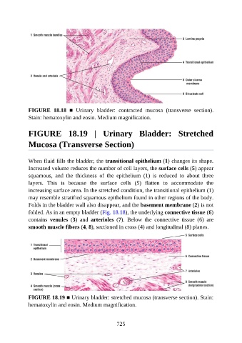
FIGURE  18.18  ■  Urinary  bladder:  contracted  mucosa  (transverse  section).
               Stain: hematoxylin and eosin. Medium magnification.


               FIGURE  18.19  |  Urinary  Bladder:  Stretched

               Mucosa (Transverse Section)


               When fluid fills the bladder, the transitional epithelium (1) changes its shape.
               Increased volume reduces the number of cell layers, the surface cells (5) appear
               squamous,  and  the  thickness  of  the  epithelium  (1)  is  reduced  to  about  three

               layers.  This  is  because  the  surface  cells  (5)  flatten  to  accommodate  the
               increasing surface area. In the stretched condition, the transitional epithelium (1)
               may resemble stratified squamous epithelium found in other regions of the body.
               Folds in the bladder wall also disappear, and the basement membrane (2) is not
               folded. As in an empty bladder (Fig. 18.18), the underlying connective tissue (6)

               contains  venules  (3)  and  arterioles  (7).  Below  the  connective  tissue  (6)  are
               smooth muscle fibers (4, 8), sectioned in cross (4) and longitudinal (8) planes.


               FIGURE 18.19 ■ Urinary bladder: stretched mucosa (transverse section). Stain:
               hematoxylin and eosin. Medium magnification.


                                                          725