
Page 724 - Atlas of Histology with Functional Correlations
P. 724
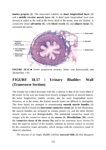
lamina propria (2).  The  muscularis  exhibits  an  inner  longitudinal  layer  (3)

               and a middle circular muscle layer (4). A third outer longitudinal layer (not
               shown) is added to the wall in the lower third of the ureter, near the bladder. A
               connective tissue adventitia (6), with blood vessels (5) and adipose tissue (7),
               surrounds the ureter.


               FIGURE  18.16  ■  Ureter  (transverse  section).  Stain:  iron  hematoxylin  and
               Alcian blue. ×10.


               FIGURE  18.17  |  Urinary  Bladder:  Wall

               (Transverse Section)


               The bladder has a thick muscular wall that is similar to that of the lower third of
               the ureter. In the wall are found three loosely arranged layers of smooth muscle,
               the  inner  longitudinal,  middle  circular,  and  the  outer  longitudinal  layers.
               However, as in the ureter, the distinct muscle layers are difficult to distinguish.

               The  three  layers  are  arranged  in  anastomosing  smooth  muscle  bundles  (1)
               between which is found the interstitial connective tissue (2). In this illustration,
               the  muscle  bundles  are  sectioned  in  various  planes  (1),  and  the  three  distinct
               muscle  layers  are  not  distinguishable.  The  interstitial  connective  tissue  (2)

               merges with the connective tissue of the serosa (3). Mesothelium (3b) covers
               the connective tissue of the serosa (3a) and is the outermost layer. Serosa (3)
               lines the superior surface of the bladder, whereas its inferior surface is covered
               by the connective tissue adventitia, which merges with the connective tissue of

               adjacent structures.

                   The mucosa of an empty bladder exhibits mucosal folds (5) that disappear


                                                          723