
Page 725 - Atlas of Histology with Functional Correlations
P. 725
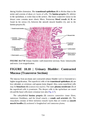
during bladder distension. The transitional epithelium (6) is thicker than in the

               ureter and consists of about six layers of cells. The lamina propria (7), inferior
               to the epithelium, is wider than in the ureters. The loose connective tissue in the
               deeper  zone  contains  more  elastic  fibers.  Numerous  blood  vessels  (4,  8)  are
               found  in  the  serosa  (3),  between  the  smooth  muscle  bundles  (1),  and  in  the
               lamina propria (8).


               FIGURE 18.17 ■ Urinary bladder: wall (transverse section). Stain: hematoxylin
               and eosin. Low magnification.


               FIGURE  18.18  |  Urinary  Bladder:  Contracted

               Mucosa (Transverse Section)


               The mucosa from an empty and contracted urinary bladder wall is illustrated at a

               higher magnification. The superficial cells of the transitional epithelium (4) are
               low cuboidal, or columnar, and appear dome shaped. Also, some superficial cells
               may be binucleate (6) (contain two nuclei). The outer plasma membrane (5) of
               the superficial cells is prominent. The deeper cells in the epithelium are round

               (4) and the basal cells more columnar (see also Fig. 4.7).

                   The  subepithelial  lamina  propria  (3)  contains  connective  tissue  fibers,
               numerous  fibroblasts,  and  the  blood  vessels,  a  venule  and  arteriole  (2).  The
               muscularis consists of three indistinct muscle layers that are visible as smooth
               muscle bundles (1) sectioned in longitudinal and transverse planes.


                                                          724