
Page 718 - Atlas of Histology with Functional Correlations
P. 718
papillary region of the kidney, and the papillary ducts (3) are spaced further
apart.
FIGURE 18.11 ■ Kidney medulla: papillary region (transverse section). Stain:
hematoxylin and eosin. Medium magnification.
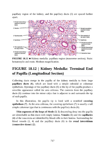
FIGURE 18.12 | Kidney Medulla: Terminal End
of Papilla (Longitudinal Section)
Collecting ducts merge in the papilla of the kidney medulla to form large
papillary ducts (6), which are lined with a simple cuboidal or columnar
epithelium. Openings of the papillary ducts (6) at the tip of the papilla produce a
sievelike appearance called the area cribrosa. The contents from the papillary
ducts (6) continue into the minor calyx that is adjacent to and surrounds the tip
of each papilla.
In this illustration, the papilla tip is lined with a stratified covering
epithelium (7). At the area cribrosa, the covering epithelium (7) is usually a tall
simple columnar type that is continuous with the papillary ducts (6).
Thin segments of the loops of Henle (3, 5) descending deep into the papilla
are identifiable as thin ducts with empty lumina. Venules (1) and the capillaries
(4) of the vasa recta are identified by blood cells in their lumina. Surrounding the
blood vessels (1, 4) and the papillary ducts (6) is the renal interstitium
(connective tissue) (2).
717