
Page 713 - Atlas of Histology with Functional Correlations
P. 713
The cells in the macula densa monitor sodium chloride concentrations in
the tubular fluid. A decrease in the blood pressure results in a decreased
glomerular filtrate and decreased sodium ion concentration in the filtrate as it
flows past the macula densa.
A decrease in systemic blood pressure or in sodium concentration in the
filtrate induces the juxtaglomerular cells to release the enzyme renin into the
bloodstream. Renin, in turn, converts the blood plasma protein
angiotensinogen to angiotensin I, which, in turn, is converted to
angiotensin II by angiotensin-converting enzyme located in the endothelial
cells of lung capillaries. Angiotensin II is an active hormone and a powerful
vasoconstrictor that initially produces arterial constriction, thereby
increasing the systemic blood pressure. In addition, angiotensin II stimulates
the release of the hormone aldosterone from the zona glomerulosa of the
adrenal gland cortex.
Aldosterone influences some cells of distal convoluted tubules, but
mainly the cells of the collecting ducts to increase their reabsorption of
sodium and chloride ions from the glomerular filtrate. Water follows sodium
chloride by osmosis and increases fluid volume in the circulatory system
raising the systemic blood pressure, increasing glomerular filtration rate, and
decreasing the secretion of renin by juxtaglomerular cells. Aldosterone also
facilitates the elimination of potassium and hydrogen ions and is an essential
hormone for maintaining electrolyte balance in the body.
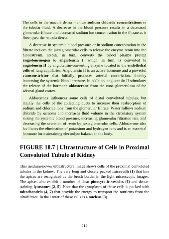
FIGURE 18.7 | Ultrastructure of Cells in Proximal
Convoluted Tubule of Kidney
This medium-power ultrastructure image shows cells of the proximal convoluted
tubules in the kidney. The very long and closely packed microvilli (1) that line
the apices are recognized as the brush border in the light microscopic images.
The apices also exhibit a number of clear pinocytotic vesicles (6) and dense-
staining lysosomes (2, 5). Note that the cytoplasm of these cells is packed with
mitochondria (4, 7) that provide the energy to transport the nutrients from the
ultrafiltrate. In the center of these cells is a nucleus (3).
712