
Page 710 - Atlas of Histology with Functional Correlations
P. 710
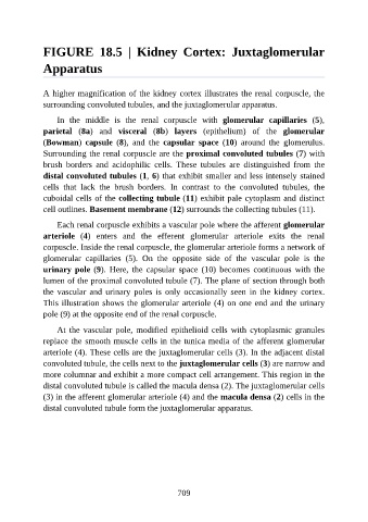
FIGURE  18.5  |  Kidney  Cortex:  Juxtaglomerular

               Apparatus


               A higher magnification of the kidney cortex illustrates the renal corpuscle, the
               surrounding convoluted tubules, and the juxtaglomerular apparatus.


                   In  the  middle  is  the  renal  corpuscle  with  glomerular  capillaries  (5),
               parietal  (8a)  and  visceral  (8b)  layers  (epithelium)  of  the  glomerular
               (Bowman)  capsule  (8),  and  the  capsular  space  (10)  around  the  glomerulus.
               Surrounding the renal corpuscle are the proximal convoluted tubules (7) with
               brush  borders  and  acidophilic  cells.  These  tubules  are  distinguished  from  the

               distal convoluted tubules (1, 6) that exhibit smaller and less intensely stained
               cells  that  lack  the  brush  borders.  In  contrast  to  the  convoluted  tubules,  the
               cuboidal cells of the collecting tubule (11) exhibit pale cytoplasm and distinct

               cell outlines. Basement membrane (12) surrounds the collecting tubules (11).

                   Each renal corpuscle exhibits a vascular pole where the afferent glomerular
               arteriole  (4)  enters  and  the  efferent  glomerular  arteriole  exits  the  renal
               corpuscle. Inside the renal corpuscle, the glomerular arteriole forms a network of
               glomerular  capillaries  (5).  On  the  opposite  side  of  the  vascular  pole  is  the

               urinary pole (9).  Here,  the  capsular  space  (10)  becomes  continuous  with  the
               lumen of the proximal convoluted tubule (7). The plane of section through both
               the  vascular  and  urinary  poles  is  only  occasionally  seen  in  the  kidney  cortex.
               This illustration shows the glomerular arteriole (4) on one end and the urinary

               pole (9) at the opposite end of the renal corpuscle.

                   At  the  vascular  pole,  modified  epithelioid  cells  with  cytoplasmic  granules
               replace the smooth muscle cells in the tunica media of the afferent glomerular
               arteriole (4). These cells are the juxtaglomerular cells (3). In the adjacent distal

               convoluted tubule, the cells next to the juxtaglomerular cells (3) are narrow and
               more columnar and exhibit a more compact cell arrangement. This region in the
               distal convoluted tubule is called the macula densa (2). The juxtaglomerular cells
               (3) in the afferent glomerular arteriole (4) and the macula densa (2) cells in the

               distal convoluted tubule form the juxtaglomerular apparatus.


                                                          709