
Page 711 - Atlas of Histology with Functional Correlations
P. 711
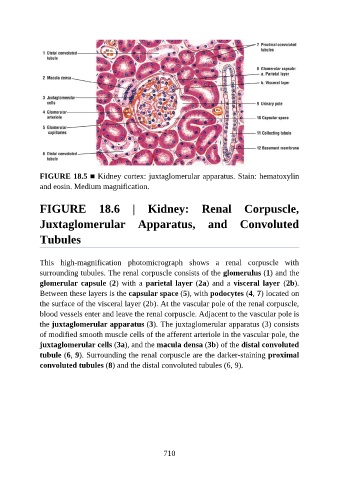
FIGURE 18.5 ■ Kidney cortex: juxtaglomerular apparatus. Stain: hematoxylin
               and eosin. Medium magnification.


               FIGURE  18.6  |  Kidney:  Renal  Corpuscle,

               Juxtaglomerular  Apparatus,  and  Convoluted


               Tubules


               This  high-magnification  photomicrograph  shows  a  renal  corpuscle  with
               surrounding tubules. The renal corpuscle consists of the glomerulus (1) and the
               glomerular capsule (2) with a parietal layer (2a) and a visceral layer  (2b).

               Between these layers is the capsular space (5), with podocytes (4, 7) located on
               the surface of the visceral layer (2b). At the vascular pole of the renal corpuscle,
               blood vessels enter and leave the renal corpuscle. Adjacent to the vascular pole is
               the juxtaglomerular apparatus (3). The juxtaglomerular apparatus (3) consists

               of modified smooth muscle cells of the afferent arteriole in the vascular pole, the
               juxtaglomerular cells (3a), and the macula densa (3b) of the distal convoluted
               tubule (6, 9). Surrounding the renal corpuscle are the darker-staining proximal
               convoluted tubules (8) and the distal convoluted tubules (6, 9).


                                                          710