
Page 712 - Atlas of Histology with Functional Correlations
P. 712
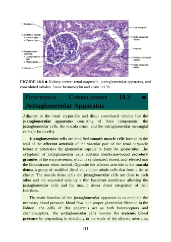
FIGURE 18.6 ■ Kidney cortex: renal corpuscle, juxtaglomerular apparatus, and
               convoluted tubules. Stain: hematoxylin and eosin. ×130.


                  FUNCTIONAL                     CORRELATIONS                       18.2            ■


                  Juxtaglomerular Apparatus


                 Adjacent  to  the  renal  corpuscles  and  distal  convoluted  tubules  lies  the
                 juxtaglomerular  apparatus  consisting  of  three  components:  the
                 juxtaglomerular cells, the macula densa, and the extraglomerular mesangial
                 cells (or lacis cells).


                     Juxtaglomerular cells are modified smooth muscle cells located in the
                 wall  of  the  afferent  arteriole  of  the  vascular  pole  of  the  renal  corpuscle
                 before  it  penetrates  the  glomerular  capsule  to  form  the  glomerulus.  The
                 cytoplasm  of  juxtaglomerular  cells  contains  membrane-bound  secretory

                 granules of the enzyme renin, which is synthesized, stored, and released into
                 the bloodstream when needed. Opposite the afferent arteriole is the macula
                 densa, a group of modified distal convoluted tubule cells that form a dense
                 cluster. The macula densa cells and juxtaglomerular cells are close to each

                 other  and  are  separated  only  by  a  thin  basement  membrane  allowing  the
                 juxtaglomerular  cells  and  the  macula  densa  closer  integration  of  their
                 functions.

                     The  main  function  of  the  juxtaglomerular  apparatus  is  to  maintain  the

                 necessary blood pressure, blood flow, and proper glomerular filtration in the
                 kidney.  The  cells  of  this  apparatus  act  as  both  baroreceptors  and
                 chemoreceptors.  The  juxtaglomerular  cells  monitor  the  systemic  blood
                 pressure by responding to stretching in the walls of the afferent arterioles.


                                                          711