
Page 715 - Atlas of Histology with Functional Correlations
P. 715
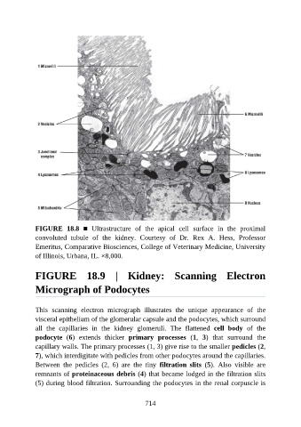
FIGURE  18.8  ■  Ultrastructure  of  the  apical  cell  surface  in  the  proximal
               convoluted  tubule  of  the  kidney.  Courtesy  of  Dr.  Rex  A.  Hess,  Professor
               Emeritus, Comparative Biosciences, College of Veterinary Medicine, University
               of Illinois, Urbana, IL. ×8,000.


               FIGURE  18.9  |  Kidney:  Scanning  Electron

               Micrograph of Podocytes


               This  scanning  electron  micrograph  illustrates  the  unique  appearance  of  the
               visceral epithelium of the glomerular capsule and the podocytes, which surround
               all  the  capillaries  in  the  kidney  glomeruli.  The  flattened  cell  body  of  the
               podocyte  (6)  extends  thicker  primary  processes  (1,  3)  that  surround  the

               capillary walls. The primary processes (1, 3) give rise to the smaller pedicles (2,
               7), which interdigitate with pedicles from other podocytes around the capillaries.
               Between  the  pedicles  (2,  6)  are  the  tiny  filtration  slits  (5).  Also  visible  are

               remnants of proteinaceous debris (4) that became lodged in the filtration slits
               (5) during blood filtration. Surrounding the podocytes in the renal corpuscle is


                                                          714