
Page 717 - Atlas of Histology with Functional Correlations
P. 717
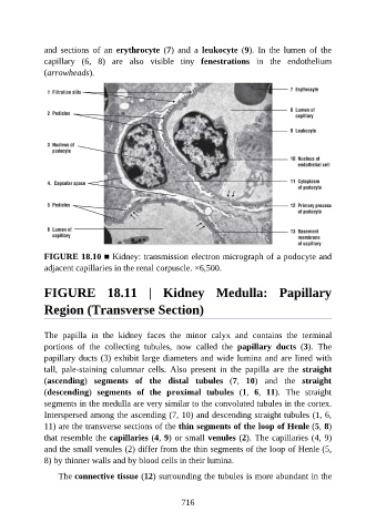
and  sections  of  an  erythrocyte  (7)  and  a  leukocyte  (9).  In  the  lumen  of  the

               capillary  (6,  8)  are  also  visible  tiny  fenestrations  in  the  endothelium
               (arrowheads).


               FIGURE 18.10 ■ Kidney: transmission electron micrograph of a podocyte and
               adjacent capillaries in the renal corpuscle. ×6,500.


               FIGURE  18.11  |  Kidney  Medulla:  Papillary

               Region (Transverse Section)


               The  papilla  in  the  kidney  faces  the  minor  calyx  and  contains  the  terminal
               portions  of  the  collecting  tubules,  now  called  the  papillary  ducts  (3).  The
               papillary ducts (3) exhibit large diameters and wide lumina and are lined with
               tall,  pale-staining  columnar  cells.  Also  present  in  the  papilla  are  the  straight

               (ascending)  segments  of  the  distal  tubules  (7,  10)  and  the  straight
               (descending)  segments  of  the  proximal  tubules  (1,  6,  11).  The  straight
               segments in the medulla are very similar to the convoluted tubules in the cortex.
               Interspersed among the ascending (7, 10) and descending straight tubules (1, 6,

               11) are the transverse sections of the thin segments of the loop of Henle (5, 8)
               that resemble the capillaries (4, 9) or small venules (2). The capillaries (4, 9)
               and the small venules (2) differ from the thin segments of the loop of Henle (5,
               8) by thinner walls and by blood cells in their lumina.


                   The connective tissue (12) surrounding the tubules is more abundant in the


                                                          716