
Page 716 - Atlas of Histology with Functional Correlations
P. 716
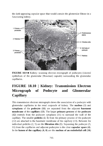
the dark-appearing capsular space that would contain the glomerular filtrate in a

               functioning kidney.


               FIGURE 18.9 ■ Kidney: scanning electron micrograph of podocytes (visceral
               epithelium  of  the  glomerular  [Bowman]  capsule)  surrounding  the  glomerular
               capillaries.


               FIGURE  18.10  |  Kidney:  Transmission  Electron

               Micrograph                 of       Podocyte              and         Glomerular


               Capillary


               This transmission electron micrograph shows the association of a podocyte with
               glomerular  capillaries  in  the  renal  corpuscle  of  kidney.  The  nucleus  (3)  and

               cytoplasm  of  the  podocyte  (11)  are  separated  from  the  adjacent  basement
               membrane of the capillary (13). The larger primary process of the podocyte
               (12)  extends  from  the  podocyte  cytoplasm  (11)  to  surround  the  wall  of  the
               capillary. The smaller pedicles (2, 5) from the primary process of the podocyte
               (12) are attached to the basement membrane of the capillary (13). Between the

               individual pedicles (2, 5) are the filtration slits (1). Separating the podocyte (3,
               11) from the capillaries and adjacent podocytes is the clear capsular space (4).
               In the lumen of the capillary (6, 8) are the nucleus of an endothelial cell (10)


                                                          715