
Page 714 - Atlas of Histology with Functional Correlations
P. 714
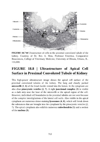
FIGURE 18.7 ■ Ultrastructure of cells in the proximal convoluted tubule of the

               kidney.  Courtesy  of  Dr.  Rex  A.  Hess,  Professor  Emeritus,  Comparative
               Biosciences, College of Veterinary Medicine, University of Illinois, Urbana, IL.
               ×55,000.


               FIGURE  18.8  |  Ultrastructure  of  Apical  Cell

               Surface in Proximal Convoluted Tubule of Kidney


               This  high-power  ultrastructure  image  shows  the  apical  cell  surface  of  the
               proximal  convoluted  tubules  of  the  kidney.  The  long  and  closely  packed
               microvilli (1, 6) of the brush border extend into the lumen. In the cytoplasm are
               also clear pinocytotic vesicles (2, 7). A tight junctional complex (3) is visible

               as a dark strip near the base of the microvilli or the apical region of the cell.
               However, individual cell boundaries in the proximal tubules are not seen because
               of the complex interdigitations of the lateral cell walls. Also visible in the apical
               cytoplasm are numerous dense-staining lysosomes (4, 8), which will break down

               the substances that are brought into the cytoplasm by the pinocytotic vesicles (2,
               7). The apical cytoplasm also exhibits numerous mitochondria (5) and a section
               of the nucleus (9).


                                                          713