
Page 889 - Atlas of Histology with Functional Correlations
P. 889
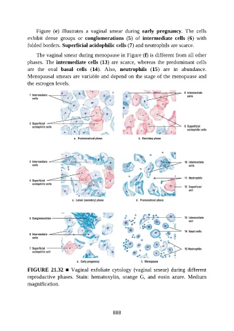
Figure  (e)  illustrates  a  vaginal  smear  during  early  pregnancy.  The  cells

               exhibit  dense  groups  or  conglomerations  (5)  of  intermediate  cells  (6)  with
               folded borders. Superficial acidophilic cells (7) and neutrophils are scarce.

                   The vaginal smear during menopause in Figure (f) is different from all other
               phases. The intermediate cells (13) are scarce, whereas the predominant cells
               are  the  oval  basal  cells  (14).  Also,  neutrophils  (15)  are  in  abundance.

               Menopausal smears are variable and depend on the stage of the menopause and
               the estrogen levels.


               FIGURE 21.32 ■ Vaginal exfoliate cytology (vaginal smear) during different
               reproductive  phases.  Stain:  hematoxylin,  orange  G,  and  eosin  azure.  Medium

               magnification.


                                                          888