
Page 893 - Atlas of Histology with Functional Correlations
P. 893
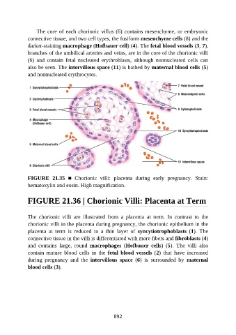
The core of each chorionic villus (6) contains mesenchyme, or embryonic

               connective tissue, and two cell types, the fusiform mesenchyme cells (8) and the
               darker-staining macrophage (Hofbauer cell) (4). The fetal blood vessels (3, 7),
               branches of the umbilical arteries and veins, are in the core of the chorionic villi
               (6)  and  contain  fetal  nucleated  erythroblasts,  although  nonnucleated  cells  can
               also be seen. The intervillous space (11) is bathed by maternal blood cells (5)

               and nonnucleated erythrocytes.


               FIGURE  21.35  ■  Chorionic  villi:  placenta  during  early  pregnancy.  Stain:

               hematoxylin and eosin. High magnification.


               FIGURE 21.36 | Chorionic Villi: Placenta at Term


               The  chorionic  villi  are  illustrated  from  a  placenta  at  term.  In  contrast  to  the
               chorionic villi in the placenta during pregnancy, the chorionic epithelium in the
               placenta  at  term  is  reduced  to  a  thin  layer  of  syncytiotrophoblasts  (1).  The
               connective tissue in the villi is differentiated with more fibers and fibroblasts (4)
               and  contains  large,  round  macrophages  (Hofbauer  cells)  (5).  The  villi  also

               contain  mature  blood  cells  in  the  fetal  blood  vessels  (2)  that  have  increased
               during  pregnancy  and  the  intervillous  space  (6)  is  surrounded  by  maternal
               blood cells (3).


                                                          892