
Page 898 - Atlas of Histology with Functional Correlations
P. 898
Activation and Early Development
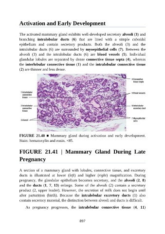
The activated mammary gland exhibits well-developed secretory alveoli (3) and
branching intralobular ducts (6) that are lined with a simple cuboidal
epithelium and contain secretory products. Both the alveoli (3) and the
intralobular ducts (6) are surrounded by myoepithelial cells (7). Between the
alveoli (3) and the intralobular ducts (6) are blood vessels (5). Individual
glandular lobules are separated by dense connective tissue septa (4), whereas
the interlobular connective tissue (1) and the intralobular connective tissue
(2) are thinner and less dense.
FIGURE 21.40 ■ Mammary gland during activation and early development.
Stain: hematoxylin and eosin. ×85.
FIGURE 21.41 | Mammary Gland During Late
Pregnancy
A section of a mammary gland with lobules, connective tissue, and excretory
ducts is illustrated at lower (left) and higher (right) magnification. During
pregnancy, the glandular epithelium becomes secretory, and the alveoli (2, 8)
and the ducts (1, 7, 13) enlarge. Some of the alveoli (2) contain a secretory
product (2, upper leader). However, the secretion of milk does not begin until
after parturition (birth). Because the intralobular excretory ducts (1) also
contain secretory material, the distinction between alveoli and ducts is difficult.
As pregnancy progresses, the intralobular connective tissue (4, 11)
897