
Page 900 - Atlas of Histology with Functional Correlations
P. 900
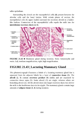
taller epithelium.

                   Surrounding the alveoli are the myoepithelial cells (8) present between the

               alveolar  cells  and  the  basal  lamina.  With  certain  planes  of  section,  the
               myoepithelial cells (8, upper leader) surround the secretory alveoli in a basket-
               like  fashion.  Contraction  of  the  myoepithelial  cells  expels  the  milk  into  the
               interlobular excretory ducts (2, 6).


               FIGURE  21.42  ■  Mammary  gland  during  lactation.  Stain:  hematoxylin  and

               eosin. Left, medium magnification; right, high magnification.


               FIGURE 21.43 | Lactating Mammary Gland


               This photomicrograph illustrates a lobule of a lactating mammary gland that is
               separated  from  the  adjacent  lobule  by  a  layer  of  connective  tissue  (5).  The
               alveoli  (2,  3)  contain  secretory  product  (6)  (milk)  and  are  separated  by
               connective  tissue  septa  (5).  Some  alveoli  (3)  are  single,  whereas  others  are
               branching alveoli (2). All the alveoli drain into larger excretory ducts that deliver

               the milk to the lactiferous ducts in the nipple. The mammary glands contain large
               amounts of adipose tissue (1, 4) during lactation.


                                                          899