
Page 901 - Atlas of Histology with Functional Correlations
P. 901
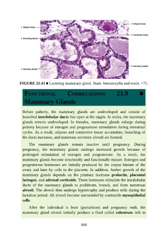
FIGURE 21.43 ■ Lactating mammary gland. Stain: hematoxylin and eosin. ×75.


                  FUNCTIONAL                     CORRELATIONS                       21.9            ■


                  Mammary Glands


                 Before  puberty,  the  mammary  glands  are  undeveloped  and  consist  of
                 branched interlobular ducts that open at the nipple. In males, the mammary
                 glands  remain  undeveloped.  In  females,  mammary  glands  enlarge  during

                 puberty because of estrogen and progesterone stimulation during menstrual
                 cycles. As a result, adipose and connective tissue accumulate, branching of
                 the ducts increases, and numerous secretory alveoli are formed.

                     The  mammary  glands  remain  inactive  until  pregnancy.  During
                 pregnancy,  the  mammary  glands  undergo  increased  growth  because  of

                 prolonged  stimulation  of  estrogen  and  progesterone.  As  a  result,  the
                 mammary glands become structurally and functionally mature. Estrogen and
                 progesterone  hormones  are  initially  produced  by  the  corpus  luteum  of  the
                 ovary  and  later  by  cells  in  the  placenta.  In  addition,  further  growth  of  the

                 mammary  glands  depends  on  the  pituitary  hormone  prolactin,  placental
                 lactogen, and adrenal corticoids. These hormones stimulate the intralobular
                 ducts  of  the  mammary  glands  to  proliferate,  branch,  and  form  numerous
                 alveoli. The alveoli then undergo hypertrophy and produce milk during the

                 lactation period. All alveoli become surrounded by contractile myoepithelial
                 cells.

                     After  the  individual  is  born  (parturition)  and  pregnancy  ends,  the
                 mammary  gland  alveoli  initially  produce  a  fluid  called  colostrum  rich  in


                                                          900