
Page 899 - Atlas of Histology with Functional Correlations
P. 899
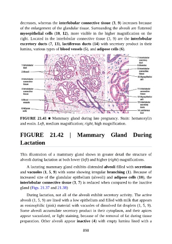
decreases, whereas the interlobular connective tissue (3, 9) increases because

               of the enlargement of the glandular tissue. Surrounding the alveoli are flattened
               myoepithelial  cells  (10,  12),  more  visible  in  the  higher  magnification  on  the
               right. Located in the interlobular connective tissue (3, 9) are the interlobular
               excretory ducts (7, 13), lactiferous ducts (14) with secretory product in their
               lumina, various types of blood vessels (5), and adipose cells (6).


               FIGURE 21.41 ■ Mammary gland during late pregnancy. Stain:  hematoxylin

               and eosin. Left, medium magnification; right, high magnification.


               FIGURE  21.42  |  Mammary  Gland  During


               Lactation


               This  illustration  of  a  mammary  gland  shows  in  greater  detail  the  structure  of
               alveoli during lactation at both lower (left) and higher (right) magnifications.

                   A lactating mammary gland exhibits distended alveoli filled with secretions

               and vacuoles (1, 5, 9) with some showing irregular branching (1). Because of
               increased size of the glandular epithelium (alveoli) and adipose cells (10), the
               interlobular connective tissue (3, 7) is reduced when compared to the inactive
               gland (Figs. 21.37 and 21.38)

                   During lactation, not all of the alveoli exhibit secretory activity. The active

               alveoli (1, 5, 9) are lined with a low epithelium and filled with milk that appears
               as eosinophilic (pink) material with vacuoles of dissolved fat droplets (1, 5, 9).
               Some alveoli accumulate secretory product in their cytoplasm, and their apices
               appear vacuolated, or light staining, because of the removal of fat during tissue

               preparation. Other alveoli appear inactive (4) with empty lumina lined with a


                                                          898