
Page 897 - Atlas of Histology with Functional Correlations
P. 897
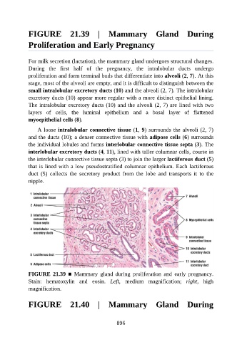
FIGURE  21.39  |  Mammary  Gland  During

               Proliferation and Early Pregnancy


               For milk secretion (lactation), the mammary gland undergoes structural changes.
               During  the  first  half  of  the  pregnancy,  the  intralobular  ducts  undergo

               proliferation and form terminal buds that differentiate into alveoli (2, 7). At this
               stage, most of the alveoli are empty, and it is difficult to distinguish between the
               small intralobular excretory ducts (10) and the alveoli (2, 7). The intralobular
               excretory ducts (10) appear more regular with a more distinct epithelial lining.

               The intralobular excretory ducts (10) and the alveoli (2, 7) are lined with two
               layers  of  cells,  the  luminal  epithelium  and  a  basal  layer  of  flattened
               myoepithelial cells (8).

                   A loose intralobular connective tissue (1, 9) surrounds the alveoli (2, 7)

               and the ducts (10); a denser connective tissue with adipose cells (6) surrounds
               the individual lobules and forms interlobular connective tissue septa (3). The
               interlobular excretory ducts (4, 11), lined with taller columnar cells, course in
               the interlobular connective tissue septa (3) to join the larger lactiferous duct (5)
               that is lined with a low pseudostratified columnar epithelium. Each lactiferous

               duct  (5)  collects  the  secretory  product  from  the  lobe  and  transports  it  to  the
               nipple.


               FIGURE  21.39  ■  Mammary  gland  during  proliferation  and  early  pregnancy.
               Stain:  hematoxylin  and  eosin.  Left,  medium  magnification;  right,  high
               magnification.


               FIGURE  21.40  |  Mammary  Gland  During


                                                          896