
Page 892 - Atlas of Histology with Functional Correlations
P. 892
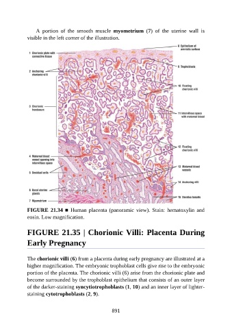
A  portion  of  the  smooth  muscle  myometrium  (7)  of  the  uterine  wall  is

               visible in the left corner of the illustration.


               FIGURE 21.34 ■  Human  placenta  (panoramic  view).  Stain:  hematoxylin  and

               eosin. Low magnification.


               FIGURE 21.35 | Chorionic Villi: Placenta During


               Early Pregnancy


               The chorionic villi (6) from a placenta during early pregnancy are illustrated at a
               higher magnification. The embryonic trophoblast cells give rise to the embryonic

               portion of the placenta. The chorionic villi (6) arise from the chorionic plate and
               become surrounded by the trophoblast epithelium that consists of an outer layer
               of the darker-staining syncytiotrophoblasts (1, 10) and an inner layer of lighter-
               staining cytotrophoblasts (2, 9).


                                                          891