
Page 891 - Atlas of Histology with Functional Correlations
P. 891
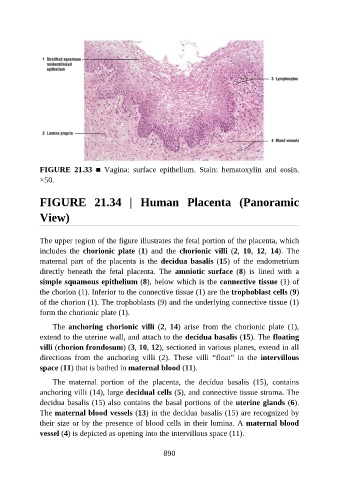
FIGURE  21.33  ■  Vagina:  surface  epithelium.  Stain:  hematoxylin  and  eosin.

               ×50.


               FIGURE  21.34  |  Human  Placenta  (Panoramic


               View)


               The upper region of the figure illustrates the fetal portion of the placenta, which
               includes  the  chorionic  plate  (1)  and  the  chorionic  villi  (2,  10,  12,  14).  The

               maternal  part  of  the  placenta  is  the  decidua basalis (15)  of  the  endometrium
               directly  beneath  the  fetal  placenta.  The  amniotic  surface  (8)  is  lined  with  a
               simple squamous epithelium (8), below which is the connective tissue (1) of
               the chorion (1). Inferior to the connective tissue (1) are the trophoblast cells (9)

               of the chorion (1). The trophoblasts (9) and the underlying connective tissue (1)
               form the chorionic plate (1).

                   The  anchoring  chorionic  villi  (2,  14)  arise  from  the  chorionic  plate  (1),
               extend to the uterine wall, and attach to the decidua basalis (15). The floating

               villi (chorion frondosum) (3, 10, 12), sectioned in various planes, extend in all
               directions  from  the  anchoring  villi  (2).  These  villi  “float”  in  the  intervillous
               space (11) that is bathed in maternal blood (11).

                   The  maternal  portion  of  the  placenta,  the  decidua  basalis  (15),  contains
               anchoring villi (14), large decidual cells (5), and connective tissue stroma. The

               decidua basalis (15) also contains the basal portions of the uterine glands (6).
               The maternal blood vessels (13) in the decidua basalis (15) are recognized by
               their size or by the presence of blood cells in their lumina. A maternal blood
               vessel (4) is depicted as opening into the intervillous space (11).


                                                          890