
Page 944 - Atlas of Histology with Functional Correlations
P. 944
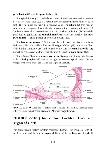
spiral lamina (2) near the spiral limbus (1).

                   The spiral limbus (1) is a thickened mass of periosteal connective tissue of

               the osseous spiral lamina (2) that extends into and forms the floor of the cochlear
               duct  (9).  The  spiral  limbus  (1)  is  covered  by  an  epithelium  (5)  that  appears
               columnar and is supported by a lateral extension of the osseous spiral lamina (2).
               The lateral extracellular extension of the spiral limbus epithelium (5) beyond the

               spiral  limbus  (1)  forms  the  tectorial  membrane  (10)  that  overlies  the  inner
               spiral tunnel (8) and a portion of the organ of Corti (13).

                   The basilar membrane (16) is a vascularized connective tissue that forms
               the lower wall of the cochlear duct (9). The organ of Corti (13) rests on the fibers

               of the basilar membrane (16) and consists of the sensory outer hair cells (11),
               supporting cells, associated inner spiral tunnel (8), and an inner tunnel (12).

                   The afferent fibers of the cochlear nerve (4) from the bipolar cells located
               in  the  spiral  ganglion  (3)  course  through  the  osseous  spiral  lamina  (2)  and
               synapse with outer hair cells (11) in the organ of Corti (13).


               FIGURE 22.17 ■ Inner ear: cochlear duct (scala media) and the hearing organ
               of Corti. Stain: hematoxylin and eosin. Medium magnification.


               FIGURE  22.18  |  Inner  Ear:  Cochlear  Duct  and

               Organ of Corti


               This  higher-magnification  photomicrograph  illustrates  the  inner  ear  with  the

               cochlear canal and the hearing organ of Corti (8) in the bony cochlea (1, 9).


                                                          943