
Page 946 - Atlas of Histology with Functional Correlations
P. 946
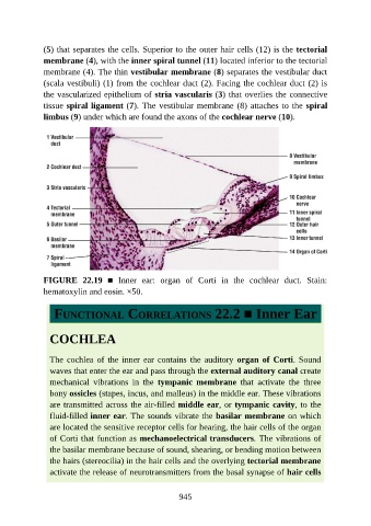
(5) that separates the cells. Superior to the outer hair cells (12) is the tectorial

               membrane (4), with the inner spiral tunnel (11) located inferior to the tectorial
               membrane (4). The thin vestibular membrane (8) separates the vestibular duct
               (scala vestibuli) (1) from the cochlear duct (2). Facing the cochlear duct (2) is
               the vascularized epithelium of stria vascularis (3) that overlies the connective
               tissue spiral ligament (7). The vestibular membrane (8) attaches to the spiral

               limbus (9) under which are found the axons of the cochlear nerve (10).


               FIGURE  22.19  ■  Inner  ear:  organ  of  Corti  in  the  cochlear  duct.  Stain:

               hematoxylin and eosin. ×50.


                  FUNCTIONAL CORRELATIONS 22.2 ■ Inner Ear


                 COCHLEA


                 The  cochlea  of  the  inner  ear  contains  the  auditory  organ  of  Corti. Sound

                 waves that enter the ear and pass through the external auditory canal create
                 mechanical  vibrations  in  the  tympanic  membrane  that  activate  the  three
                 bony ossicles (stapes, incus, and malleus) in the middle ear. These vibrations
                 are transmitted across the air-filled middle ear, or tympanic cavity, to the
                 fluid-filled inner ear. The sounds vibrate the basilar membrane on which

                 are located the sensitive receptor cells for hearing, the hair cells of the organ
                 of Corti that function as mechanoelectrical transducers. The vibrations of
                 the basilar membrane because of sound, shearing, or bending motion between

                 the hairs (stereocilia) in the hair cells and the overlying tectorial membrane
                 activate the release of neurotransmitters from the basal synapse of hair cells


                                                          945