
Page 943 - Atlas of Histology with Functional Correlations
P. 943
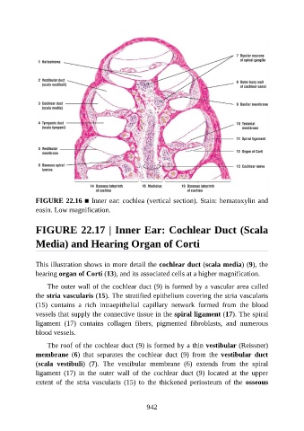
FIGURE 22.16 ■ Inner ear: cochlea (vertical section). Stain: hematoxylin and

               eosin. Low magnification.


               FIGURE 22.17 | Inner Ear: Cochlear Duct (Scala


               Media) and Hearing Organ of Corti


               This illustration shows in more detail the cochlear duct (scala media) (9), the
               hearing organ of Corti (13), and its associated cells at a higher magnification.

                   The outer wall of the cochlear duct (9) is formed by a vascular area called

               the stria vascularis (15). The stratified epithelium covering the stria vascularis
               (15)  contains  a  rich  intraepithelial  capillary  network  formed  from  the  blood
               vessels that supply the connective tissue in the spiral ligament (17). The spiral
               ligament  (17)  contains  collagen  fibers,  pigmented  fibroblasts,  and  numerous
               blood vessels.


                   The roof of the cochlear duct (9) is formed by a thin vestibular (Reissner)
               membrane  (6)  that  separates  the  cochlear  duct  (9)  from  the  vestibular  duct
               (scala  vestibuli)  (7).  The  vestibular  membrane  (6)  extends  from  the  spiral
               ligament  (17)  in  the  outer  wall  of  the  cochlear  duct  (9)  located  at  the  upper

               extent  of  the  stria  vascularis  (15)  to  the  thickened  periosteum  of  the  osseous


                                                          942