
Page 945 - Atlas of Histology with Functional Correlations
P. 945
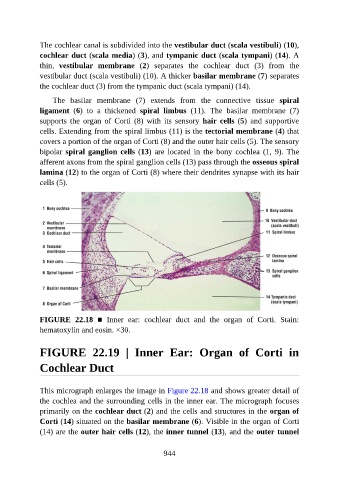
The cochlear canal is subdivided into the vestibular duct (scala vestibuli) (10),

               cochlear duct (scala media) (3), and tympanic duct (scala tympani) (14). A
               thin,  vestibular  membrane  (2)  separates  the  cochlear  duct  (3)  from  the
               vestibular duct (scala vestibuli) (10). A thicker basilar membrane (7) separates
               the cochlear duct (3) from the tympanic duct (scala tympani) (14).

                   The  basilar  membrane  (7)  extends  from  the  connective  tissue  spiral

               ligament  (6)  to  a  thickened  spiral  limbus  (11).  The  basilar  membrane  (7)
               supports  the  organ  of  Corti  (8)  with  its  sensory  hair cells (5)  and  supportive
               cells. Extending from the spiral limbus (11) is the tectorial membrane (4) that
               covers a portion of the organ of Corti (8) and the outer hair cells (5). The sensory

               bipolar spiral ganglion cells (13)  are  located  in  the  bony  cochlea  (1,  9).  The
               afferent axons from the spiral ganglion cells (13) pass through the osseous spiral
               lamina (12) to the organ of Corti (8) where their dendrites synapse with its hair
               cells (5).


               FIGURE  22.18  ■  Inner  ear:  cochlear  duct  and  the  organ  of  Corti.  Stain:

               hematoxylin and eosin. ×30.


               FIGURE  22.19  |  Inner  Ear:  Organ  of  Corti  in


               Cochlear Duct


               This micrograph enlarges the image in Figure 22.18 and shows greater detail of
               the cochlea and the surrounding cells in the inner ear. The micrograph focuses

               primarily on the cochlear duct (2) and the cells and structures in the organ of
               Corti (14) situated on the basilar membrane (6). Visible in the organ of Corti
               (14) are the outer hair cells (12), the inner tunnel (13), and the outer tunnel


                                                          944