
Page 941 - Atlas of Histology with Functional Correlations
P. 941
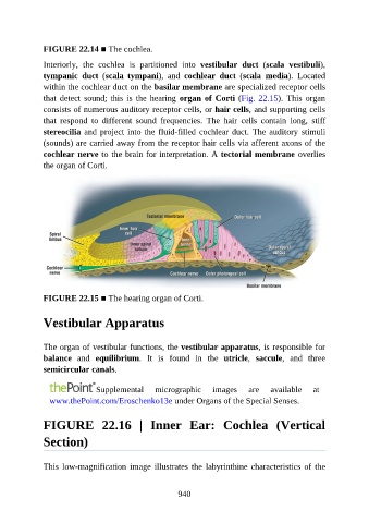
FIGURE 22.14 ■ The cochlea.

               Interiorly,  the  cochlea  is  partitioned  into  vestibular  duct  (scala  vestibuli),

               tympanic  duct  (scala  tympani),  and  cochlear  duct  (scala  media).  Located
               within the cochlear duct on the basilar membrane are specialized receptor cells
               that  detect  sound;  this  is  the  hearing  organ of Corti (Fig. 22.15).  This  organ
               consists of numerous auditory receptor cells, or hair cells, and supporting cells

               that  respond  to  different  sound  frequencies.  The  hair  cells  contain  long,  stiff
               stereocilia and project into the fluid-filled cochlear duct. The auditory stimuli
               (sounds) are carried away from the receptor hair cells via afferent axons of the
               cochlear nerve to the brain for interpretation. A tectorial membrane overlies

               the organ of Corti.


               FIGURE 22.15 ■ The hearing organ of Corti.


               Vestibular Apparatus


               The organ of vestibular functions, the vestibular apparatus, is responsible for
               balance  and  equilibrium.  It  is  found  in  the  utricle,  saccule,  and  three

               semicircular canals.


                                Supplemental       micrographic       images     are    available     at
                 www.thePoint.com/Eroschenko13e under Organs of the Special Senses.


               FIGURE  22.16  |  Inner  Ear:  Cochlea  (Vertical

               Section)


               This  low-magnification  image  illustrates  the  labyrinthine  characteristics  of  the


                                                          940