
Page 47 - DP Vol 19 No 2 HR_Neat
P. 47
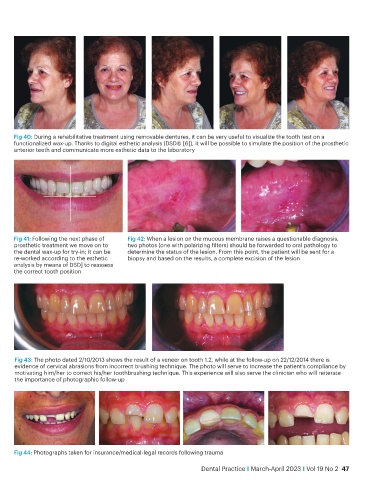
Fig 40: During a rehabilitative treatment using removable dentures, it can be very useful to visualize the tooth test on a
           functionalized wax-up. Thanks to digital esthetic analysis (DSD© [6]), it will be possible to simulate the position of the prosthetic
           anterior teeth and communicate more esthetic data to the laboratory


           Fig 41: Following the next phase of   Fig 42: When a lesion on the mucous membrane raises a questionable diagnosis,
           prosthetic treatment we move on to   two photos (one with polarizing filters) should be forwarded to oral pathology to
           the dental wax-up for try-in; it can be   determine the status of the lesion. From this point, the patient will be sent for a
           re-worked according to the esthetic   biopsy and based on the results, a complete excision of the lesion
           analysis by means of DSD] to reassess
           the correct tooth position


            Fig 43: The photo dated 2/10/2013 shows the result of a veneer on tooth 1.2, while at the follow-up on 22/12/2014 there is
            evidence of cervical abrasions from incorrect brushing technique. The photo will serve to increase the patient’s compliance by
            motivating him/her to correct his/her toothbrushing technique. This experience will also serve the clinician who will reiterate
            the importance of photographic follow-up


           Fig 44: Photographs taken for insurance/medical-legal records following trauma

                                                                         Dental Practice i March-April 2023 i Vol 19 No 2  47