
Page 48 - DP Vol 20 No 5_Neat
P. 48
ORTHODONTIC SECTION
ORTHODONTIC CORRECTION OF LUCY G
Rhona Eskander describes a case she completed of a patient complaining of crooked laterals
and uneven occlusal surfaces
Throughout this article we will look at a case treated under the principles of
the SAFE orthodontics education program. The methodology is based on a
checklist of four main principles:
1. Stability
2. Assessment
3. Function
4. Aesthetics
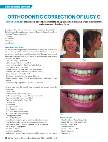
PATIENT COMPLAINT
The patient came complaining because she felt as though her laterals 'stuck
out and the edges of the front teeth are uneven'. She wanted to align the
upper teeth to match the upper right one. Round out the upper arch to match
the present position of UR6/UL6. Finish with an overset of 2-4mm. During
checkup, it was noted:
• Lower face height – decreased. Fig 1 Fig 2
• Upper midline to facial – coincident
• Lower midline to facial – slightly shifted to the left.
• Chin point to facial – coincident
• Face profile view – skeletal class 2 and severity mild
• Smile analysis – high smile line and smile arc convex.
• Buccal corridors – mildly deficient
• Tooth shape normal with some incisal chipping
• Tooth size and symmetry - laterals are small also due to inadequate gingival
zeniths.
• Soft tissues – lip competent, no lip catch, NLA normal.
However, one must be careful with alignment top ensure proper lip Fig 3
competence.
• Overjet 2mm
• Incisor classification – class II div II
• Molar relationship right – class II three-quarter unit
• Canine relationship right – class II full
• Canine relationship left – class II full.
• Overbite 30%
• No cross bite
• No displacements
• Crowding upper 0-4mm
• Arch form upper – square shape
• Arch form lower – square shape Fig 4
• Oral hygiene excellent
• No dental pathologies. PROBLEM LIST
• Lateral excursion right – group function and no interferences 1. Class II molar on the right hand side
• Lateral excursion left – group function and no interferences. 2. Class II full unit canine on both side
• Protrusive excursion – anterior guidance and no interference 3. Chipped anterior teeth
• TMD - no pain on palpation 4. Discoloured teeth
• No clicks or crepitus 5. Lowered centre-line shift
• Mouth opening 40mm. 6. Class two incisors
• Radiograph – no pathology, with generalized horizontal bone loss 1-2mm. 7. Thin biotype
48 Dental Practice I November-December 2024 I Vol 20 No 5