
Page 505 - Atlas of Small Animal CT and MRI
P. 505
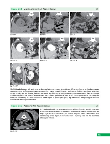
Figure 5.1.6  Migrating Foreign Body Abscess (Canine)                                       CT


            (a) CT, TP                       (b) CT, TP                       (c) CT, TP


            (d) CT+C, TP                     (e) CT+C, TP                     (f) CT+C, TP


            (g) CT+C, DP
            12y FS Labrador Retriever with acute onset of abdominal pain, recent history of coughing, and fever. Unenhanced (a–c) and comparable
            contrast‐enhanced (d–f) transverse images are ordered from cranial to caudal. There is a well‐circumscribed tract and abscess in the right
            retroperitoneal space lateral to the diaphragmatic muscle (d,g: black arrow) with peripheral contrast enhancement. There is additional
            nonenhancing fluid dorsal to the inflammatory tract, which contains gas bubbles (d: open arrow). The retroperitoneal fat surrounding the
            kidney has fat stranding in a linear pattern with central regions of ground‐glass opacity (f,g: arrowheads). A migrating grass awn was
            retrieved from the retroperitoneal space.


              Figure 5.1.7  Abdominal Wall Abscess (Canine)                                               CT
                                             8y FS Border Collie with a recurrent abscess on the left flank. There is a multilobulated mass
                                             in the subcutaneous tissues of the left dorsolateral body wall. The mass does not enter the
                                             deeper layers of fat adjacent to the spine. There is peripheral contrast enhancement with
                                             nonenhancing central regions. Plant material from a migrating grass awn was discovered
                                             after surgical removal.


            (a) CT+C, TP
                                                                                                             495