
Page 510 - Atlas of Small Animal CT and MRI
P. 510
500 Atlas of Small Animal CT and MRI
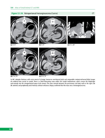
Figure 5.1.16 Retroperitoneal Hemangiosarcoma (Canine) CT
(a) CT, TP (b) CT, TP (c) CT+C, DP
(d) CT+C, TP (e) CT+C, TP
3y MC Labrador Retriever with acute onset of lethargy. Transverse unenhanced (a,b) and comparable contrastenhanced (d,e) images
are ordered from cranial to caudal. There is a fluid‐attenuating mass within the caudal mediastinum, which crosses the diaphragm
and extends into the retroperitoneal space (c,d: arrows). The mass extends between the peritoneum and body wall on the right side
(d: asterisk) and peripherally and intensely contrast enhances. Biopsy confirmed that the mass was a hemangiosarcoma.
500