
Page 511 - Atlas of Small Animal CT and MRI
P. 511
Body Wall, Retroperitoneum, and Peritoneal Cavity 501
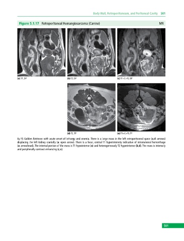
Figure 5.1.17 Retroperitoneal Hemangiosarcoma (Canine) MR
(a) T1, DP (b) T2, DP (c) T1+C+FS, DP
(d) T2, TP (e) T1+C+FS, TP
6y FS Golden Retriever with acute onset of lethargy and anemia. There is a large mass in the left retroperitoneal space (a,d: arrows)
displacing the left kidney cranially (a: open arrow). There is a focal, central T1 hyperintensity indicative of intratumoral hemorrhage
(a: arrowhead). The internal portion of the mass is T1 hypointense (a) and heterogeneously T2 hyperintense (b,d). The mass is intensely
and peripherally contrast enhancing (c,e).
501