
Page 512 - Atlas of Small Animal CT and MRI
P. 512
502 Atlas of Small Animal CT and MRI
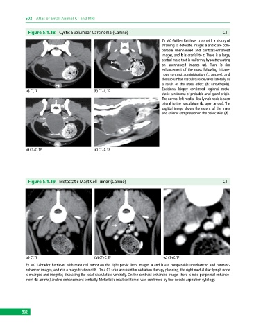
Figure 5.1.18 Cystic Sublumbar Carcinoma (Canine) CT
7y MC Golden Retriever cross with a history of
straining to defecate. Images a and c are com
parable unenhanced and contrastenhanced
images, and b is cranial to c. There is a large,
central mass that is uniformly hypoattenuating
on unenhanced images (a). There is rim
enhancement of the mass following intrave
nous contrast administration (c: arrows), and
the sublumbar vasculature deviates laterally as
a result of the mass effect (b: arrowheads).
Excisional biopsy confirmed regional meta
(a) CT, TP (b) CT+C, TP
static carcinoma of probable anal gland origin.
The normal left medial iliac lymph node is seen
lateral to the vasculature (b: open arrow). The
sagittal image shows the extent of the mass
and colonic compression in the pelvic inlet (d).
(c) CT+C, TP (d) CT+C, SP
Figure 5.1.19 Metastatic Mast Cell Tumor (Canine) CT
(a) CT, TP (b) CT+C, TP (c) CT+C, TP
7y MC Labrador Retriever with mast cell tumor on the right pelvic limb. Images a and b are comparable unenhanced and contrast
enhanced images, and c is a magnification of b. On a CT scan acquired for radiation therapy planning, the right medial iliac lymph node
is enlarged and irregular, displacing the local vasculature ventrally. On the contrast‐enhanced image, there is mild peripheral enhance
ment (b: arrows) and no enhancement centrally. Metastatic mast cell tumor was confirmed by fine‐needle aspiration cytology.
502