
Page 516 - Atlas of Small Animal CT and MRI
P. 516
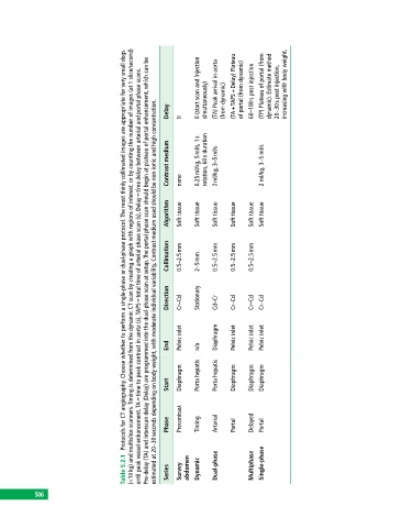
(<10 kg) and multislice scanners. Timing is determined from the dynamic CT scan by creating a graph with regions of interest, or by counting the number of images (at 1 slice/second)
                                       Protocols for CT angiography. Choose whether to perform a single‐phase or dual‐phase protocol. The most thinly collimated images are appropriate for very small dogs
                                                             0 (start scan and injection   (TA) Peak arrival in aorta    (TA +  TAPS + Delay) Plateau   of portal (from dynamic)  60–180 s post injection  (TP) Plateau of portal (from  dynamic). Estimate method   20–30 s post injection,  increasing with body weight.
                                             Pre‐delay (TA) and interscan delay (Delay) are programmed into the dual‐phase scan at setup. The portal phase scan should begin at plateau of portal enhancement, which can be
                                           until peak vessel enhancement. TA = time to peak contrast in aorta (s), TAPS = total time of arterial phase scan (s), Delay = time delay between arterial and portal phase scans.


                                                estimated at 20–30 seconds depending on body weight, with moderate individual variability. Contrast medium used should be non‐ionic and high concentration.
                                                    Delay  0   simultaneously)  (from dynamic)


                                                    Contrast medium  none  0.25 ml/kg, 5 ml/s, 1 s   rotation, 60 s duration  2 ml/kg, 3–5 ml/s  2 ml/kg, 3–5 ml/s


                                                    Algorithm  Soft tissue  Soft tissue  Soft tissue  Soft tissue  Soft tissue  Soft tissue


                                                    Collimation  0.5–2.5 mm  2–5 mm  0.5–2.5 mm  0.5–2.5 mm  0.5–2.5 mm


                                                    Direction  Cr–Cd  Stationary  Cd–Cr  Cr–Cd  Cr–Cd  Cr–Cd


                                                       Pelvic inlet  Diaphragm  Pelvic inlet  Pelvic inlet  Pelvic inlet

                                                    End      n/a


                                                    Start  Diaphragm  Porta hepatis  Porta hepatis  Diaphragm  Diaphragm  Diaphragm


                                                    Phase  Precontrast  Timing  Arterial  Portal  Delayed  Portal


                                       Table 5.2.1   Series  Survey   abdomen  Dynamic  Dual‐phase  Multiphase  Single‐phase


           506