
Page 521 - Atlas of Small Animal CT and MRI
P. 521
Hepatovascular Disorders 511
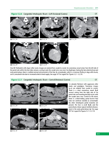
Figure 5.2.6 Congenital Intrahepatic Shunt—Left Divisional (Canine) CT
(a) CT+C, TP (b) CT+C, TP (c) CT+C, TP
(d) CT+C, TP (e) CT+C, TP
3mo MC Rottweiler with stupor after meals. Images are ordered from caudal to cranial. An anomalous vessel arises from the left side of
the portal vein within the liver (d,e: arrows), curving to join the caudal vena cava near the diaphragm. During the late arterial phase and
early portal phase, there is mottled contrast enhancement of the liver (b: arrowheads), which is a common finding in dogs with shunts,
and is presumed to be due to increased arterial blood supply. See page 507 for Legend for Figures 5.2.1–5.2.18.
Figure 5.2.7 Congenital Intrahepatic Shunt—Central Divisional (Canine) CT
1y Labrador Retriever with aggression after
meals and polydipsia. Transverse images
(a–c) are ordered from caudal to cranial.
There is a short anomalous vessel (b,d:
arrows) arising from the right side of the
portal vein and traveling dorsally to join with
the caudal vena cava. Cranial to the shunt,
there is a blind‐ending portal branch that
does not continue cranially (c: arrowhead).
No other intrahepatic portal branches are
present. The liver is small (b,d), and the
(a) CT+C, TP (b) CT+C, TP
parenchyma shows typical mottled enhance-
ment in the early portal phase (a). See page
507 for Legend for Figures 5.2.1–5.2.18.
(c) CT+C, TP (d) CT+C, SP
511