Page 523 - Atlas of Small Animal CT and MRI
P. 523

Hepatovascular Disorders  513


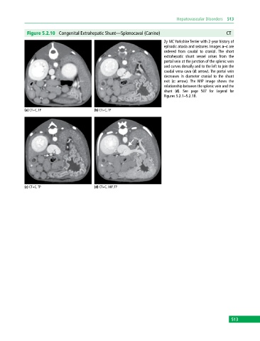
              Figure 5.2.10  Congenital Extrahepatic Shunt—Splenocaval (Canine)                           CT
                                                                             2y MC Yorkshire Terrier with 2‐year history of
                                                                             episodic ataxia and seizures. Images a–c are
                                                                             ordered  from  caudal  to  cranial.  The  short
                                                                             extrahepatic shunt  vessel arises from  the
                                                                               portal vein at the junction of the splenic vein
                                                                             and curves dorsally and to the left to join the
                                                                             caudal vena cava (d: arrow). The portal vein
                                                                             decreases  in  diameter  cranial  to  the  shunt
                                                                             exit (c: arrow).  The MIP image shows the
                                                                               relationship between the splenic vein and the
                                                                             shunt (d). See page 507 for Legend for
                                                                             Figures 5.2.1–5.2.18.


            (a) CT+C, TP                     (b) CT+C, TP


            (c) CT+C, TP                     (d) CT+C, MIP, TP


                                                                                                             513
   518   519   520   521   522   523   524   525   526   527   528