
Page 524 - Atlas of Small Animal CT and MRI
P. 524
514 Atlas of Small Animal CT and MRI
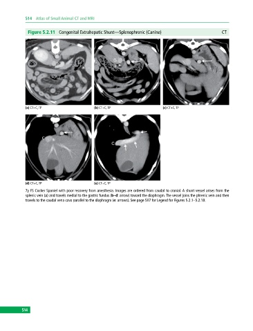
Figure 5.2.11 Congenital Extrahepatic Shunt—Splenophrenic (Canine) CT
(a) CT+C, TP (b) CT+C, TP (c) CT+C, TP
(d) CT+C, TP (e) CT+C, TP
7y FS Cocker Spaniel with poor recovery from anesthesia. Images are ordered from caudal to cranial. A shunt vessel arises from the
splenic vein (a) and travels medial to the gastric fundus (b–d: arrow) toward the diaphragm. The vessel joins the phrenic vein and then
travels to the caudal vena cava parallel to the diaphragm (e: arrows). See page 507 for Legend for Figures 5.2.1–5.2.18.
514