
Page 529 - Atlas of Small Animal CT and MRI
P. 529
Hepatovascular Disorders 519
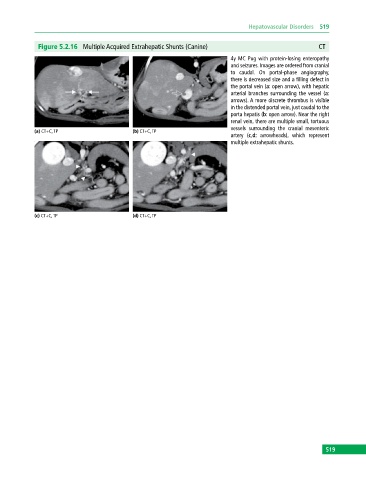
Figure 5.2.16 Multiple Acquired Extrahepatic Shunts (Canine) CT
4y MC Pug with protein‐losing enteropathy
and seizures. Images are ordered from cranial
to caudal. On portal‐phase angiography,
there is decreased size and a filling defect in
the portal vein (a: open arrow), with hepatic
arterial branches surrounding the vessel (a:
arrows). A more discrete thrombus is visible
in the distended portal vein, just caudal to the
porta hepatis (b: open arrow). Near the right
renal vein, there are multiple small, tortuous
vessels surrounding the cranial mesenteric
(a) CT+C, TP (b) CT+C, TP
artery (c,d: arrowheads), which represent
multiple extrahepatic shunts.
(c) CT+C, TP (d) CT+C, TP
519