
Page 534 - Atlas of Small Animal CT and MRI
P. 534
524 Atlas of Small Animal CT and MRI
Cysts appear as single or multiple fluid‐attenuating or Degenerative or cirrhotic livers are variably sized,
T2 hyperintense lesions with a thin wall, central fluid with chronic loss of normal parenchyma resulting in
accumulation and no enhancement (Figure 5.3.16). microhepatia. The combination of fibrosis and the lobu
When large, cysts produce a mass effect. lar abnormal architecture created by septa is termed
Degenerative disease of the liver can result in vacuolar cirrhosis. 12
hepatopathy, fat accumulation, and cyst formation,
changing the imaging characteristics of the parenchyma.
Liver disease resulting in vacuolar hepatopathy may Cholelithiasis and biliary obstruction
cause hepatic enlargement with rounded borders and Cholelithiasis, mineralized sludge accumulation, or
focal regions of hypoattenuation (Figure 5.3.17). calculi in the biliary tree may be observed in asympto
Steatosis of the liver results in more hypoattenuating matic animals (Figures 5.3.18, 5.3.19). When obstruction
parenchyma on CT images, although this may only be of the bile duct occurs, the gallbladder may be enlarged
apparent on measurements with regions of interest. with surrounding inflammation, thickened wall, and
Hepatic lipidosis in cats also results in decreased hepatic peritoneal effusion (Figure 5.3.20). Neoplasia can also
attenuation on CT images. Fat accumulation would be occur in the region of the bile duct, obstructing the flow
19
expected to be hyperintense on T1 and T2 MR images. of bile (Figure 5.3.14).
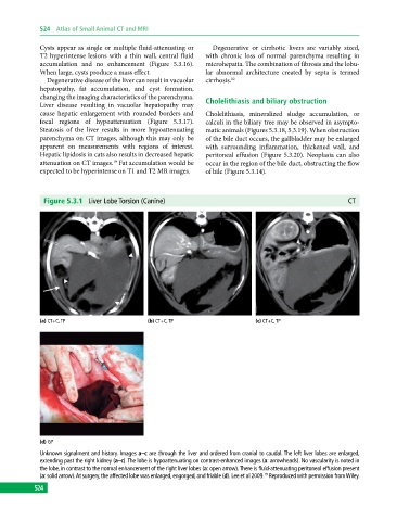
Figure 5.3.1 Liver Lobe Torsion (Canine) CT
(a) CT+C, TP (b) CT+C, TP (c) CT+C, TP
(d) GP
Unknown signalment and history. Images a–c are through the liver and ordered from cranial to caudal. The left liver lobes are enlarged,
extending past the right kidney (a–c). The lobe is hypoattenuating on contrast‐enhanced images (a: arrowheads). No vascularity is noted in
the lobe, in contrast to the normal enhancement of the right liver lobes (a: open arrow). There is fluid‐attenuating peritoneal effusion present
(a: solid arrow). At surgery, the affected lobe was enlarged, engorged, and friable (d). Lee et al 2009. Reproduced with permission from Wiley.
19
524