
Page 530 - Atlas of Small Animal CT and MRI
P. 530
520 Atlas of Small Animal CT and MRI
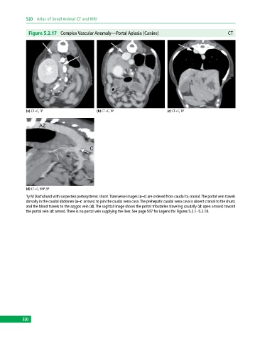
Figure 5.2.17 Complex Vascular Anomaly—Portal Aplasia (Canine) CT
(a) CT+C, TP (b) CT+C, TP (c) CT+C, TP
(d) CT+C, MIP, SP
1y M Dachshund with suspected portosystemic shunt. Transverse images (a–c) are ordered from caudal to cranial. The portal vein travels
dorsally in the caudal abdomen (a–c: arrows) to join the caudal vena cava. The prehepatic caudal vena cava is absent cranial to the shunt,
and the blood travels to the azygos vein (d). The sagittal image shows the portal tributaries traveling caudally (d: open arrows) toward
the portal vein (d: arrow). There is no portal vein supplying the liver. See page 507 for Legend for Figures 5.2.1–5.2.18.
520