Page 535 - Atlas of Small Animal CT and MRI
P. 535

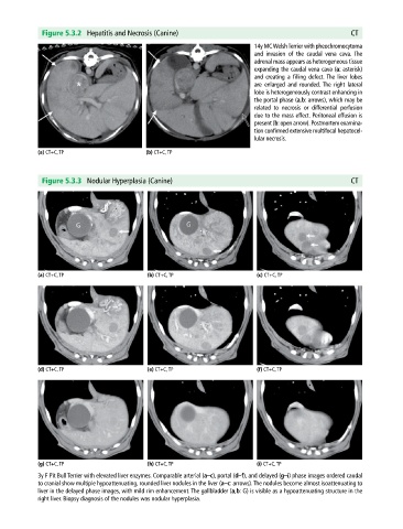
Figure 5.3.2  Hepatitis and Necrosis (Canine)                                               CT

                                                                             14y MC Welsh Terrier with pheochromocytoma
                                                                             and invasion of the caudal vena cava.  The
                                                                             adrenal mass appears as heterogeneous tissue
                                                                             expanding the caudal vena cava (a: asterisk)
                                                                             and  creating a filling defect. The liver lobes
                                                                             are  enlarged and rounded. The right   lateral
                                                                             lobe is heterogeneously contrast enhancing in
                                                                             the portal phase (a,b: arrows), which may be
                                                                             related to necrosis or differential   perfusion
                                                                             due to the mass effect. Peritoneal effusion is
                                                                             present (b: open arrow). Post mortem examina-
                                                                             tion confirmed extensive multifocal hepatocel-
                                                                             lular necrosis.

            (a) CT+C, TP                     (b) CT+C, TP


              Figure 5.3.3  Nodular Hyperplasia (Canine)                                                  CT


            (a) CT+C, TP                     (b) CT+C, TP                     (c) CT+C, TP


            (d) CT+C, TP                     (e) CT+C, TP                     (f) CT+C, TP


            (g) CT+C, TP                     (h) CT+C, TP                     (i) CT+C, TP
            3y F Pit Bull Terrier with elevated liver enzymes. Comparable arterial (a–c), portal (d–f), and delayed (g–i) phase images ordered caudal
            to cranial show multiple hypoattenuating, rounded liver nodules in the liver (a–c: arrows). The nodules become almost isoattenuating to
            liver in the delayed phase images, with mild rim enhancement. The gallbladder (a,b: G) is visible as a hypoattenuating structure in the
            right liver. Biopsy diagnosis of the nodules was nodular hyperplasia.
   530   531   532   533   534   535   536   537   538   539   540