
Page 538 - Atlas of Small Animal CT and MRI
P. 538
528 Atlas of Small Animal CT and MRI
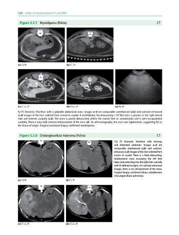
Figure 5.3.7 Myelolipoma (Feline) CT
(a) CT, TP (b) CT, TP
(c) CT+C, TP (d) CT+C, TP (e) US, SP
9y FS Domestic Shorthair with a palpable abdominal mass. Images a–d are comparable unenhanced (a,b) and contrast‐enhanced
(c,d) images of the liver ordered from cranial to caudal. A multilobular, fat‐attenuating (−67 HU) mass is present in the right lateral
lobe and extends caudally (a,b). The mass is poorly demarcated within the cranial liver (c: arrowheads) and is well encapsulated
caudally. There is very mild contrast enhancement of the mass (d). On ultrasonography, the mass was hyperechoic, suggesting fat as
the tissue of origin. Surgical excisional biopsy confirmed myelolipoma.
Figure 5.3.8 Cholangiocellular Adenoma (Feline) CT
13y FS Domestic Shorthair with lethargy
and distended abdomen. Images a–d are
comparable unenhanced (a,b) and contrast‐
enhanced (c,d) images of the liver ordered from
cranial to caudal. There is a fluid‐attenuating,
multicameral mass occupying the left liver
lobes and extending into the right liver cranially
with ill‐defined margins. On contrast‐enhanced
images, there is no enhancement of the mass.
Surgical biopsy confirmed biliary cystadenoma
(cholangiocellular adenoma).
(a) CT, TP (b) CT, TP
(c) CT+C, TP (d) CT+C, TP