
Page 541 - Atlas of Small Animal CT and MRI
P. 541
Hepatobiliary Disorders 531
Figure 5.3.12 Metastatic Hemangiosarcoma (Canine) CT
(a) CT+C, TP (b) CT+C, TP (c) CT+C, TP
(d) CT+C, TP (e) CT+C, TP (f) CT+C, TP
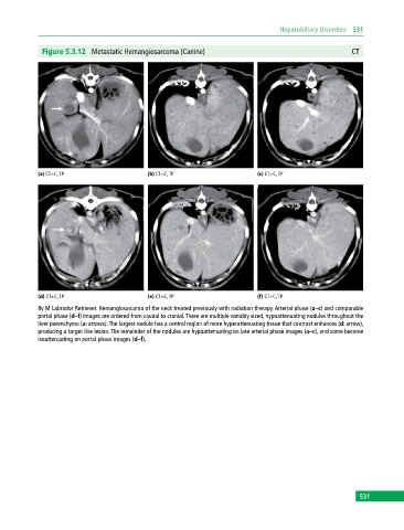
8y M Labrador Retriever. Hemangiosarcoma of the neck treated previously with radiation therapy. Arterial phase (a–c) and comparable
portal phase (d–f) images are ordered from caudal to cranial. There are multiple variably sized, hypoattenuating nodules throughout the
liver parenchyma (a: arrows). The largest nodule has a central region of more hyperattenuating tissue that contrast enhances (d: arrow),
producing a target‐like lesion. The remainder of the nodules are hypoattenuating on late arterial phase images (a–c), and some become
isoattenuating on portal phase images (d–f).
531