
Page 545 - Atlas of Small Animal CT and MRI
P. 545
Hepatobiliary Disorders 535
Figure 5.3.17 Vacuolar Hepatopathy (Canine) CT
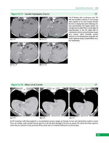
13y FS Brittany with a pulmonary mass. The
liver was included in a thoracic CT scan. Images
were acquired at the same location through
the liver at varying time points following con-
trast administration. The hepatic borders are
enlarged and rounded. There is a focal region of
hypoattenuation in the left medial lobe on
unenhanced, arterial, and portal phase images
(a–c: arrows), which minimally contrast
enhances on the delayed phase image (d). Fine
needle aspiration biopsy revealed diffuse vacu-
(a) CT, TP (b) CT+C, TP
olar hepatopathy.
(c) CT+C, TP (d) CT+C, TP
Figure 5.3.18 Biliary Calculi (Canine) CT
(a) CT, TP (b) CT, TP (c) CT, TP
4y MC Australian Cattle Dog imaged for a musculoskeletal sarcoma. Images are through the liver and ordered from caudal to cranial.
There are multiple, small, rounded mineral opacities in the bile ducts throughout the liver (a: arrows). The calculi form linear opacities
where they are clustered in close proximity. Biliary calculi were an incidental finding with no clinical signs.
535