
Page 550 - Atlas of Small Animal CT and MRI
P. 550
540 Atlas of Small Animal CT and MRI
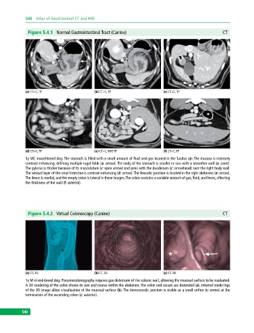
Figure 5.4.1 Normal Gastrointestinal Tract (Canine) CT
(a) CT+C, TP (b) CT+C, TP (c) CT+C, TP
(d) CT+C, TP (e) CT+C, MIP, TP (f) CT+C, TP
3y MC mixed‐breed dog. The stomach is filled with a small amount of fluid and gas located in the fundus (a). The mucosa is intensely
contrast enhancing, defining multiple rugal folds (a: arrow). The body of the stomach is smaller in size with a smoother wall (a: caret).
The pylorus is thicker because of its musculature (c: open arrow) and joins with the duodenum (c: arrowhead) near the right body wall.
The serosal layer of the small intestine is contrast enhancing (d: arrow). The ileocolic junction is located in the right abdomen (e: arrow).
The ileum is medial, and the empty colon is lateral in these images. The colon contains a variable amount of gas, fluid, and feces, affecting
the thickness of the wall (f: asterisk).
Figure 5.4.2 Virtual Colonoscopy (Canine) CT
(a) CT, 3D (b) CT, 3D (c) CT, 3D
1y M mixed‐breed dog. Pneumocolonography requires gas distension of the colonic wall, allowing the mucosal surface to be evaluated.
A 3D rendering of the colon shows its size and course within the abdomen. The colon and cecum are distended (a). Internal renderings
of the 3D image allow visualization of the mucosal surface (b). The ileocecocolic junction is visible as a small orifice (c: arrow) at the
termination of the ascending colon (c: asterisk).
540