
Page 554 - Atlas of Small Animal CT and MRI
P. 554
544 Atlas of Small Animal CT and MRI
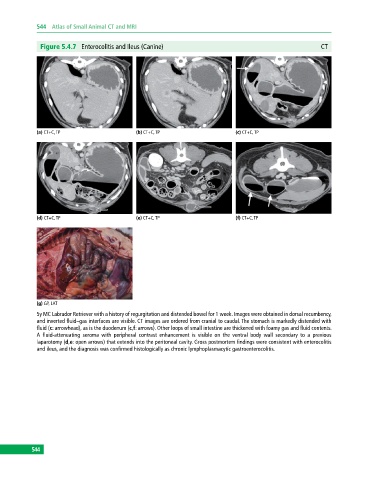
Figure 5.4.7 Enterocolitis and Ileus (Canine) CT
(a) CT+C, TP (b) CT+C, TP (c) CT+C, TP
(d) CT+C, TP (e) CT+C, TP (f) CT+C, TP
(g) GP, LAT
5y MC Labrador Retriever with a history of regurgitation and distended bowel for 1 week. Images were obtained in dorsal recumbency,
and inverted fluid–gas interfaces are visible. CT images are ordered from cranial to caudal. The stomach is markedly distended with
fluid (c: arrowhead), as is the duodenum (c,f: arrows). Other loops of small intestine are thickened with foamy gas and fluid contents.
A fluid‐attenuating seroma with peripheral contrast enhancement is visible on the ventral body wall secondary to a previous
laparotomy (d,e: open arrows) that extends into the peritoneal cavity. Gross postmortem findings were consistent with enterocolitis
and ileus, and the diagnosis was confirmed histologically as chronic lymphoplasmacytic gastroenterocolitis.
544