Page 556 - Atlas of Small Animal CT and MRI
P. 556

546  Atlas of Small Animal CT and MRI


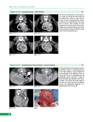
              Figure 5.4.10  Leiomyosarcoma—Colon (Feline)                                                 CT
                                                                              9y FS Domestic Mediumhair with hematoche-
                                                                              zia. Images are through the distal colon and
                                                                              are ordered from cranial to caudal. There is
                                                                              a large, eccentric, heterogeneously contrast‐
                                                                              enhancing mass originating from the colonic
                                                                              wall (a: arrows). More caudally, the mass
                                                                              compresses the colonic lumen, which is iden-
                                                                              tified by gas and mineral‐opacity feces (b–d).
                                                                              Surgical excisional biopsy confirmed a diag-
                                                                              nosis of colonic leiomyosarcoma.
             (a) CT+C, TP                     (b) CT+C, TP


             (c) CT+C, TP                     (d) CT+C, TP


              Figure 5.4.11  Gastrointestinal Stromal Tumor—Cecum (Canine)                                 CT
                                                                              7y FS Golden Retriever with progressive abdom-
                                                                              inal distension. There is an extremely large mass
                                                                              in the right abdomen, causing displacement
                                                                              of the remainder of the abdominal viscera to
                                                                              the left side. The mass is heterogeneously and
                                                                              peripherally contrast enhancing.  A region of
                                                                              bowel containing gas, identified as the cecum,
                                                                              is  visible in the dorsal and cranial aspect of
                                                                              the mass (a,c: arrow). The mass was surgically
                                                                              excised and histologically diagnosed as a
                                                                                gastrointestinal stromal tumor (d).
             (a) CT+C, TP                     (b) CT+C, TP


             (c) CT+C, DP                     (d) GP


           546
   551   552   553   554   555   556   557   558   559   560   561