
Page 557 - Atlas of Small Animal CT and MRI
P. 557
Gastrointestinal Tract 547
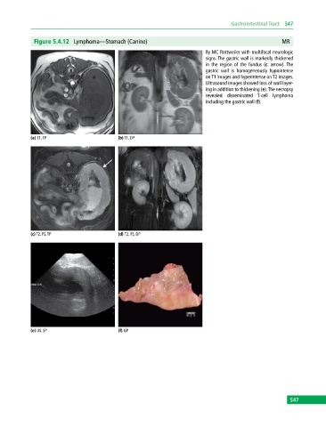
Figure 5.4.12 Lymphoma—Stomach (Canine) MR
8y MC Rottweiler with multifocal neurologic
signs. The gastric wall is markedly thickened
in the region of the fundus (c: arrow). The
gastric wall is homogeneously hypointense
on T1 images and hyperintense on T2 images.
Ultrasound images showed loss of wall layer-
ing in addition to thickening (e). The necropsy
revealed disseminated T‐cell lymphoma
including the gastric wall (f).
(a) T1, TP (b) T1, DP
(c) T2, FS, TP (d) T2, FS, DP
(e) US, SP (f) GP
547