
Page 558 - Atlas of Small Animal CT and MRI
P. 558
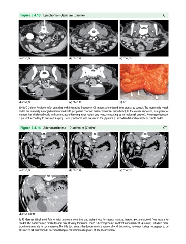
Figure 5.4.13  Lymphoma—Jejunum (Canine)                                                     CT


             (a) CT+C, TP                     (b) CT+C, TP                     (c) CT+C, TP


             (d) CT+C, TP                     (e) CT+C, TP                     (f) GP
             10y MC Golden Retriever with vomiting with increasing frequency. CT images are ordered from cranial to caudal. The mesenteric lymph
             nodes are markedly enlarged and rounded with peripheral contrast enhancement (a: arrowhead). In the caudal abdomen, a segment of
             jejunum has thickened walls with a contrast‐enhancing inner region and hypoattenuating outer region (d: arrows). Pneumoperitoneum
             is present secondary to previous surgery. T‐cell lymphoma was present in the jejunum (f: arrowheads) and mesenteric lymph nodes.


              Figure 5.4.14  Adenocarcinoma—Duodenum (Canine)                                              CT


             (a) CT+C, TP                     (b) CT+C, TP                     (c) CT+C, TP


             (d) CT+C, MIP, TP
             9y FS German Wirehaired Pointer with anorexia, vomiting, and weight loss for several months. Images a–c are ordered from cranial to
             caudal. The duodenum is markedly and eccentrically thickened. There is heterogeneous contrast enhancement (a: arrow), which is more
             prominent centrally in some regions. The bile duct enters the duodenum in a region of wall thickening; however, it does not appear to be
             obstructed (d: arrowhead). Excisional biopsy confirmed a diagnosis of adenocarcinoma.